
Illumina and Agilent Microarray Scanner Updates Signal a New Phase for High-Throughput Array Analysis
A microarray chip can contain an enormous amount of biological information, but that information is initially represented through fluorescent signals on a physical surface. The scanner converts those signals into digital images that can subsequently be analyzed for gene expression, genotyping, methylation, copy-number variation and other genomic characteristics.
The National Center for Biotechnology Information describes microarrays as solid surfaces carrying large sets of oligonucleotide probes that interact with nucleic-acid targets. Fluorescent detection then allows the signal at individual array locations to be measured.
That makes the scanner much more than an imaging peripheral. It is a critical link between biochemical hybridization and usable genomic information.
What Actually Happens Inside a Microarray Scanner?
The scanning process is built around a combination of lasers, optical focusing, detectors, motion control and image-processing electronics. Fluorescently labelled material on the array is excited by a laser, and the resulting optical signal is captured at extremely small spatial intervals.
Older technical descriptions from the U.S. National Cancer Institute explain that commercial microarray scanners commonly use confocal detection, with approximately 3-micron resolution historically being typical. The scanner measures fluorescence pixel by pixel before software converts the image into probe-level measurements.
The basic workflow can therefore be viewed as:
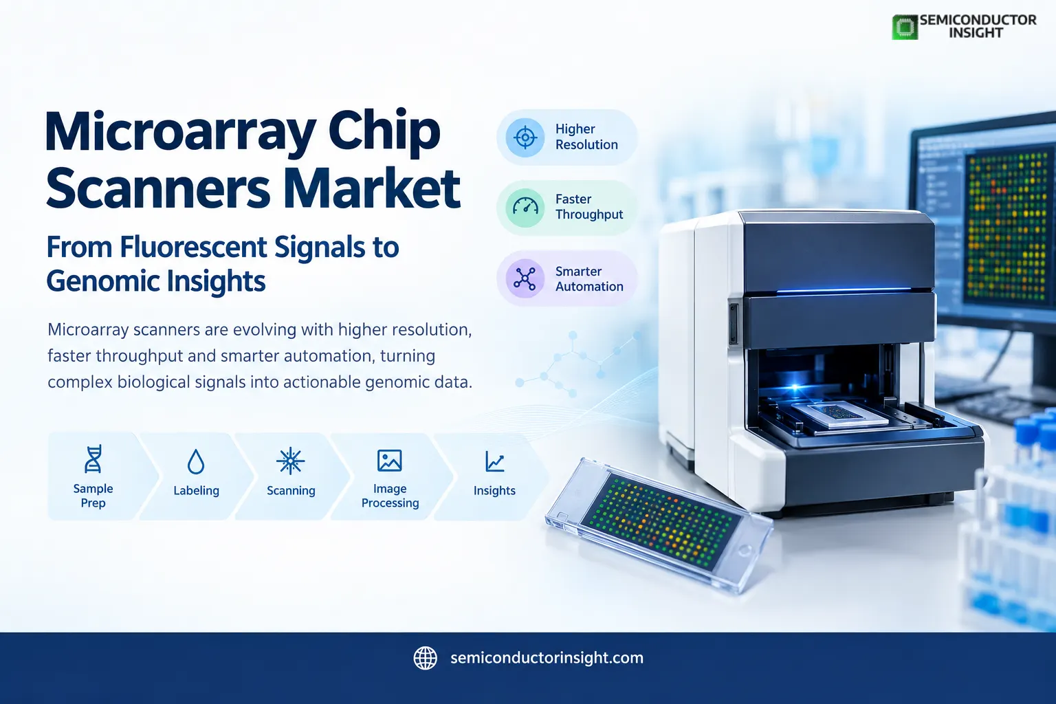
Microarray preparation → Fluorescent labeling → Laser excitation → Optical detection → Pixel generation → Image processing → Genomic interpretation
Resolution Is Becoming a Key Differentiator
- Current systems demonstrate how far scanner optics have progressed. Illumina’s iScan System specifies a pixel resolution of 0.53 μm and uses dual-laser excitation at 532 nm and 658 nm. Agilent’s SureScan platforms offer selectable scanning at 2, 3, 5 and 10 μm resolutions.
- The difference matters because smaller pixels can capture finer spatial information across dense array features. However, higher resolution also creates larger image files and can increase acquisition time, meaning laboratories have to balance optical detail with throughput.
Throughput Is Moving From Hours toward Continuous Processing
High-throughput laboratories increasingly need scanners that can process arrays without creating another bottleneck after sample preparation.
Illumina reports iScan scan times ranging from 0.3 to 26 minutes per sample depending on the array and workflow. The company says its array scanners can process thousands of samples per day.
Agilent approaches throughput differently through continuous slide loading and automated handling. Its high-resolution scanner specifications include a 48-slide autoloader, allowing laboratories to queue multiple slides rather than manually loading every individual array.
This shift is particularly relevant for population genomics, large research cohorts and laboratories processing repeated batches.
Dense Arrays Are Increasing the Optical Workload
- Modern bead-based arrays illustrate why scanner performance continues to matter.
- Illumina states that its BeadChip technology can contain hundreds of thousands to millions of genotypes for a single individual.
- The beads carry oligonucleotide probes that capture specific genomic loci, with fluorescent intensity providing information about the resulting allele signal.
- As array density increases, scanner uniformity, autofocus performance, dynamic range and signal discrimination become increasingly important.
- A scanner must distinguish useful fluorescence from background while maintaining consistent measurements across the entire slide.
The 16-Bit to 20-Bit Imaging Question
Image depth is another technical area receiving attention. Agilent’s high-resolution scanner specifications identify dynamic ranges of 10⁴ with 16-bit data, 10⁵ with 20-bit data and up to 10⁶ with extended dynamic-range scanning.
Its SureScan documentation also shows the storage implications. A 2-micron single-pass scan of a 61 × 21.6 mm region can generate approximately 1,300 MB using 16-bit TIFF output and take around 24 minutes. At 3-micron resolution, the corresponding single-pass scan is approximately 620 MB and takes about 16 minutes.
For laboratories running hundreds of arrays, these numbers quickly become a data-management consideration rather than merely an instrument specification.
To find out more, feel free to browse our latest updated report: https://semiconductorinsight.com/report/microarray-chip-scanners-market-2/
Microarray Scanning Is Expanding Across More Than One Genomic Task
The scanner’s role now extends across several established applications. Illumina identifies genotyping, DNA methylation and cytogenomic arrays among the applications supported by its array-scanning systems. Its broader microarray technology is also used for high-throughput genetic analysis.
This creates demand across research institutions, pharmaceutical development, population studies and molecular diagnostics. Agilent’s SureScan Dx system further demonstrates the movement toward regulated workflows, with the company stating that the instrument carries IVD marking in multiple markets and is manufactured under ISO 13485 certification.
The Scanner Is Becoming Part of an Automated Data Pipeline
The next development is not simply about making lasers brighter or pixels smaller. Modern systems increasingly connect scanning with automated sample handling, barcode identification, image-quality assessment and downstream analysis.
Illumina’s iScan specifications include automatic image-quality analysis and barcode-related workflow capabilities, while Agilent systems incorporate barcode reading and automated slide handling.
For high-volume laboratories, this means the Microarray Chip Scanners Market is increasingly tied to workflow automation. The competitive question is shifting from “How quickly can one slide be scanned?” toward “How efficiently can an entire batch move from chip to validated dataset?”
Where the Market Is Heading in 2026
- Microarray technology is operating alongside sequencing rather than simply disappearing because of it.
- Illumina continues to maintain dedicated array technologies while also developing sequencing and multiomics platforms.
- Its current technology portfolio explicitly positions microarrays alongside next-generation sequencing for different genetic-analysis requirements.
- That creates a distinctive opportunity for scanner manufacturers.
- High-density arrays, submicron imaging, automated loading, dual-color fluorescence, larger dynamic ranges and increasingly integrated software are turning the scanner into a sophisticated semiconductor-enabled optical system.
In 2026, the most important development is therefore not simply faster scanning. It is the convergence of optical precision, automated sample handling and high-volume genomic data generation in one increasingly integrated laboratory workflow.

Comments (0)